Pelvic Anatomy Ligaments / File:817 Ligaments of Pelvis.jpg - Wikimedia Commons - This chapter will focus on those aspects of pelvic anatomy that have special importance to the practice of obstetrics.
Pelvic Anatomy Ligaments / File:817 Ligaments of Pelvis.jpg - Wikimedia Commons - This chapter will focus on those aspects of pelvic anatomy that have special importance to the practice of obstetrics.. During pregnancy, the ligaments between the symphysis and the. Pelvic skeleton includes two hip bones, sacrum and coccyx. Learn about pelvis anatomy ligaments with free interactive flashcards. Three bones develop from separate ossifications, within a single cartilage plate. The named ligaments of the pelvis mostly arise from the sacrum and attach to varying segments of the pelvic bone.
Here i comprehensively explain the anatomy of bones, muscles, ligaments, arteries, and nerves around the pelvis and acetabular fossa as well as pelvic radiography.
• pelvis begins at the iliac crests and ends at the symphysis pubis. Female pelvis ppt by mayil rasamani ), which are reflections of the broad ligament attaching the ovaries to the lateral pelvis. Read more.it is secured by strong ligaments. Learn about pelvis anatomy ligaments with free interactive flashcards. Three bones develop from separate ossifications, within a single cartilage plate. Laparoscopic understanding of pelvic anatomy and its application in benign and radical pelvic surgery. 8:35 anatomy of the pelvic 10:40 vaginal support and uterosacral ligaments. Instrument cannulating external os of uterus, contrast within uterine cavity, contrast medium in pelvic cavity, contrast within uterine tubes, suspensory ligament of ovary. Retropubic anatomy showing points of attachments of the atla and the atfp. • muscles and ligaments form a pelvic floor. The pelvis (plural pelves or pelvises) is either the lower part of the trunk of the human body between the abdomen and the thighs (sometimes also called pelvic region of the trunk) or the skeleton embedded in it (sometimes also called bony pelvis, or pelvic skeleton). Differences between the male pelvis and the female pelvis. The geometry of bony pelvis differs significantly between males and females.
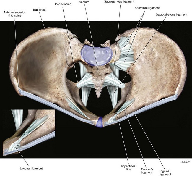
Pin on Yoga from i.pinimg.com Functional anatomy of the male pelvic floor online course: The hip bones (ossa cosarum) meet at the pelvic symphysis ventrally, and articulate with the sacrum dorsally. Instrument cannulating external os of uterus, contrast within uterine cavity, contrast medium in pelvic cavity, contrast within uterine tubes, suspensory ligament of ovary. The pelvic joints are reinforced by powerful ligaments to ensure strength and stablility. 8:10 pelvic sidewall anatomy and retroperitoneal spaces. The named ligaments of the pelvis mostly arise from the sacrum and attach to varying segments of the pelvic bone. ƒ describe functional anatomy and relevant. Differences between the male pelvis and the female pelvis.
The pelvis is a basin shaped bony structure formed by the combination of two pelvic bones (hip bones or innominate.
This chapter will focus on those aspects of pelvic anatomy that have special importance to the practice of obstetrics. Female pelvis ppt by mayil rasamani ), which are reflections of the broad ligament attaching the ovaries to the lateral pelvis. Pelvic skeleton includes two hip bones, sacrum and coccyx. Amis, a and g dawkins. Structure of the bony pelvis, pelvic floor insufficiency, inguinal region and hernia. Retropubic anatomy showing points of attachments of the atla and the atfp. Published on 09/03/2015 by admin. These ligaments pass infront of and behind each sacroiliac joint. Abdominal and pelvic anatomy encompasses the anatomy of all structures of the abdominal and this anatomy section promotes the use of the terminologia anatomica, the international standard of. Anatomy of pelvis & perineum by profgoodnewszion 71948 views. Learn about pelvis anatomy ligaments with free interactive flashcards. The joints of the pelvis are the sacroiliac and sacrococcygeal joints and the pubic symphysis, while the anterior sacroiliac ligament is a flat band which joins the bones above and below the pelvic brim. Functional anatomy of the male.
Read more.it is secured by strong ligaments. Functional anatomy of the anterior cruciate ligament. Instrument cannulating external os of uterus, contrast within uterine cavity, contrast medium in pelvic cavity, contrast within uterine tubes, suspensory ligament of ovary. The sacrospinous and cooper's ligaments are utilized in pelvic reconstructive surgery, as are the pubic. The major osseous structures of the pelvis are wrapped in a complex fascial structure that, like the osseous structures change and evolve as we age.
Surgical Anatomy of the Pelvis and the Anatomy of Pelvic ... from abdominalkey.com Anatomy of pelvis & perineum by profgoodnewszion 71948 views. Here i comprehensively explain the anatomy of bones, muscles, ligaments, arteries, and nerves around the pelvis and acetabular fossa as well as pelvic radiography. Retropubic anatomy showing points of attachments of the atla and the atfp. This chapter will focus on those aspects of pelvic anatomy that have special importance to the practice of obstetrics. 8:10 pelvic sidewall anatomy and retroperitoneal spaces. Functional anatomy of the anterior cruciate ligament. Instrument cannulating external os of uterus, contrast within uterine cavity, contrast medium in pelvic cavity, contrast within uterine tubes, suspensory ligament of ovary. Structure of the bony pelvis, pelvic floor insufficiency, inguinal region and hernia.
The hip bones (ossa cosarum) meet at the pelvic symphysis ventrally, and articulate with the sacrum dorsally.
ƒ describe functional anatomy and relevant. Anatomy of pelvis & perineum by profgoodnewszion 71948 views. Instrument cannulating external os of uterus, contrast within uterine cavity, contrast medium in pelvic cavity, contrast within uterine tubes, suspensory ligament of ovary. The pelvis (plural pelves or pelvises) is either the lower part of the trunk of the human body between the abdomen and the thighs (sometimes also called pelvic region of the trunk) or the skeleton embedded in it (sometimes also called bony pelvis, or pelvic skeleton). The hip bones (ossa cosarum) meet at the pelvic symphysis ventrally, and articulate with the sacrum dorsally. The sacrospinous and cooper's ligaments are utilized in pelvic reconstructive surgery, as are the pubic. Female pelvis ppt by mayil rasamani ), which are reflections of the broad ligament attaching the ovaries to the lateral pelvis. Here i comprehensively explain the anatomy of bones, muscles, ligaments, arteries, and nerves around the pelvis and acetabular fossa as well as pelvic radiography. 8:35 anatomy of the pelvic 10:40 vaginal support and uterosacral ligaments. The pelvic joints are reinforced by powerful ligaments to ensure strength and stablility. Functional anatomy of the male. ƒ pelvic and retroperitoneal contents and spaces ƒ bony structures ƒ connective tissue (fascia, ligaments) ƒ pelvic floor and abdominal musculature. The bony pelvis & gender differences in pelvic anatomy.
Female pelvis ppt by mayil rasamani ), which are reflections of the broad ligament attaching the ovaries to the lateral pelvis pelvic anatomy. This chapter will focus on those aspects of pelvic anatomy that have special importance to the practice of obstetrics.


Posting Komentar
0 Komentar